Chowdhury Rafiqul Ahsan
Department of Microbiology, Dhaka University, Dhaka, Bangladesh.
Kohinur Begum
Department of Pharmacy, ASA University, Dhaka, Bangladesh.
Enamul Kabir
Department of Pathology, Sir Salimullah Medical College, Dhaka, Bangladesh.
Alam Nur-E-Kamal
Department of Biology, Medgar Evers College of the City University of New York, Brollklyn, N.Y. 11225, USA
Kaisar Ali Talukder
Department of Biotechnology and Genetic Engineering, Mawlana Bhashani Science and Technology University, Tangail, Bangladesh.
Keywords: Stx2d, cytotoxic effect, enterotoxic effect, neurotoxic effect
DOI: 10.3329/bmrcb.v46i1.47468
Abstract
Background: Shiga toxin (Stx) producing Escherichia coli (STEC) colonize human intestinal tract and their infections have asymptomatic clinical manifestations which cause local and systemic pathological changes.
Objectives: This study intended to establish the role of Shiga toxin (Stx2d) in developing clinical manifestations in STEC infections using experimental models.
Methods: A total 300 stool samples were screened from hospitalised diarrhoeal patients enrolled in 2% surveillance system at International Centre for Diarrhoeal Disease Research, Bangladesh (icddr,b). The stx gene profile including their variants was identified byPCR.stx2d gene positive STEC PT187 was selected for toxin (s) preparation.Toxin was prepared by centrifugation of culture supernatant. Enterotoxicand paralytic-lethal activities were tested in rabbit ileal loops and mice, respectively. Histopathological study of the rabbit ileal loop segments and different tissues of mice by paraffin embedded method and stained by H & E staining. Cytotoxic effect was performed on HeLa cells.
Results: Nine STEC strains were identified for stx2 gene positive. Among them STEC PT187 was found stx2d gene positive strain and selected for toxic activities. Toxin (s) responsible for causing accumulation of fluid in rabbit ileal loops and its segments showed inflammation and enterocytenecrosis. In mouse model, toxin(s) was found to cause hind limbs paralysis and death. Brain, spinal cord and kidney tissue of mice showed histopathological changes. Toxin (s) also showed positive cytotoxic activity in HeLa cell.
Conclusions: In this study, results indicated that Stx2d producing >E. coli exhibit not only enterotoxic activity, but also cause impaired neurological functions and cytotoxic effect.
Keywords: Stx2d, cytotoxic effect, enterotoxic effect, neurotoxic effect.Introduction
Shiga toxin producing Escherichia coli (STEC) strains have emerged as one of the majorcause of human gastrointestinal diseases resulting in life-threatening complications such as hemolytic-uremic syndrome (HUS) which is the most common cause of acute renal failure in children. In some cases, patients infected by STEC, also showed neurological symptoms.1 Epidemiological studies from different parts of the world established STEC as the major cause of watery or bloody diarrhea and HUS,mostly in temperate climatic regions.2,3 Domestic animals, mainly cattle, sheep, and goats, have been established as major natural reservoirs for STEC and play a significant role in the epidemiology of human infections. In most of the developing countries, hygienic conditions are severely compromised and living with domestic animals within the same premises is a common practice. Although the socioeconomic status and living style of the people in South Asian countries including Bangladesh support the prevalence of STEC infections, however, only few studies have been conducted in this region until now. This is probably because the STEC isnot considered as a common pathogen among hospitalized patients in Bangladesh suffering from mild cases of diarrhoea within the community.2On the other hand, development of therapeutic and preventative strategies to combat STEC infections requires a thorough understanding of the mechanisms by which STEC organisms colonize the human intestinal tract and cause local and systemic pathological changes. Pathogenicity of STEC is associated with their ability to produce diverse virulence and toxic factors. The main factors are the ability to produce shiga toxins which can be subdivided into shiga toxin1 (Stx1) and shiga toxin2 (Stx2). Nucleotide sequence analysis of the stx1 and stx2 genes revealed the existence of different variants in both groups. So far, three stx1 subtypes (stx1, stx1c, stx1d) and several stx2 variants (e.g. stx2c, stx2d, stx2e, stx2f, stx2g) have been described.4-7
Among these variants, Stx2 showed the strongest toxic effects and Stx2d has displayed cytoxicityto Vero cells, which increased by 10-1000 folds.4,8 Several studies have shown that the prevalence of strains having stx2dgene is present more in livestock reservoirs than in humans. Stx2d was found to be significantly associated with bloody diarrhoea and systemic complications such as HUS.9 On the other hand, shigatoxin is also found neurotoxic and destroyed neurons.10 However, molecular basis of neuronal damage caused by the shiga toxin has remained unclear. Aim of this study to investigate the role of shiga toxintype 2d produced by E.coli, in producing enterotoxic, neurotoxic and cytotoxic effects using animal models and mammalian cell culture.
Materials and Methods
Ethics statement: The 2.0% surveillance system was a routine ongoing activity of the icddr,b Dhaka Hospital which has been approved by the Research Review Committee (RRC) and Ethical Review Committee (ERC) of icddr,b. Since most of the patients were illiterate, informed oral consent was obtained from the caregivers or guardians on behalf of the patients for collecting stool specimens only, following the hospital policy. The information was stored in the hospital database and used for conducting research. The verbal consent was documented by keeping a checkmark in the questionnaire which was again shown to the patient or the guardians. At the same time, patients or the guardians were assured about the non-disclosure of information collected from them, and were also informed about the use of data for analysis and using the results for improving patient
Isolation and identification of STEC strain: A total of 300 stool specimens collected from icddr,bdiarrhoea treatment centre, under the systematic surveillance system from 2.0% patients attending at the icddr,b diarrhoea treatment centre, between December 2001 and August 2002, every fifth patient attending the hospital was included for sampling. Stool samples were collected in sterile stool container, were examined for STECstrainisolation. All samples were tested for STECisolation following standard methods,11,12 by PCR using stx1and stx2primers with their variants.5,6,13-15 Isolated STECstrains were reconfirmed for stx1, stx2and stx2 variants according to the protocol described.16 Isolatedstrains were serologically confirmed by using commercially available antisera kit. Preparation of toxin(s):Toxin(s) was prepared following the method described,17,18 briefly, strain was grown aerobically at 37 °C in Trypticase Soy Broth (TSB) for 24 h and culture was centrifuged (13,000 g for 20 min) and supernatant was concentrated (1 mg/ml) over sucrose.19 Concentrated supernatant was filtered through 0.22 mm millipore membrane. Protein concentration of the sample was estimated by Bio-Rad protein assay according to Bradford method.
Rabbit ileal loop assay: Enterotoxic effect was performed in New Zealand rabbitsas described by Singh et al,.20 1 ml of toxin(s) was inoculated into rabbit ileal loops. V. cholerae 569B and VTEC were used as positive controls. Sterile TSB was used as negative control. Each test was done using two rabbits. The length of each loop and the volume of fluid accumulated were measured to determine the amount of fluid accumulation per unit length of gut (ml/cm). After rabbit ileal loop assay,histopathological study of full-thickness segments of the ligated ileum was performed by paraffin embedded method and stained by haematoxylin and eosin (H&E) staining and examined under light microscope.The histological changes were graded from 0 to 4+, with 0 (normal) being no change and 4+ equalling severe inflammation andenterocytic necrosis.21
RITARD model test:Ten ml of cell culture suspension in PBS (3x10⁸ CFU/ml) was injected into the lumen of the anterior jejunum of adult New Zealand rabbits.22 Each test was done using two rabbits. Rabbits were observed for signs and symptoms of diarrhoea, weakness and death. Rectal swabs were taken daily and plated onto MacConkey agar to identify shedding of the test organism. Isolated colonies were confirmed by PCR analysis.Ten ml PBS was injected in duplicate rabbits were used as negative controls.
Mouse lethality test:Five groups of Swiss Albino mice (six per group), 6 to 8 weeks old,were injected intra-peritoneally (IP) with five different doses (0.1µg, 1µg, 5µg, 10µg, 50µg per mice) of toxin(s).TSB was used as negative control. Deaths were monitored twice daily and 50.0% lethal dose (LD50) was determined.23 Mice were observed for signs of toxic effect until death. Histopathological study of brain, spinal cord and kidney tissues were performed according to the procedures described earlier.
Cytotoxic assay:Cytotoxic effect of toxin(s) in HeLa cell linewasdetermined.24 Morphological alteration of the HeLa cell was observed after staining the actin filaments with fluorescein isothiocyanate (FITC)–conjugated phalloidinunder epifluorescence microscope.25
Results
Isolation and identification of STEC strainsby PCR: Nine strainsout of 300 stoolsamples were identifiedfor STEC specific genestx2 (tableI). Among them only one representative STEC(PT187 stx2dgene positive) strain was selected for this study.This strain was further analysed for serological typing and confirmed to belong to the O2 serotype.
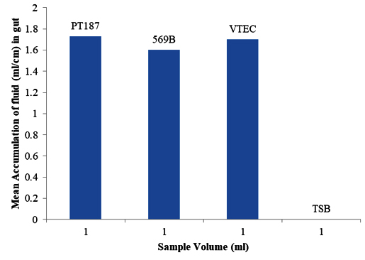
Induction of fluid accumulation in rabbit ileal loop: Toxin(s) of STEC PT187 caused fluid accumulationin rabbit ileal loops. V. cholerae 569B and VTEC were found to cause accumulation of fluid, whileTSB (negative control) did not (Figure 1).
| Sample no | stx1>gene | stx>2 gene |
| PT49 | - | + |
| PT92 | - | + |
| PT149 | - | + |
| PT164 | + | + |
| PT187 | - | + |
| PT210 | - | + |
| PT271 | - | + |
| PT277 | - | + |
| PT281 | - | + |
| VTEC3 | + | + |
| K-12 | - | - |
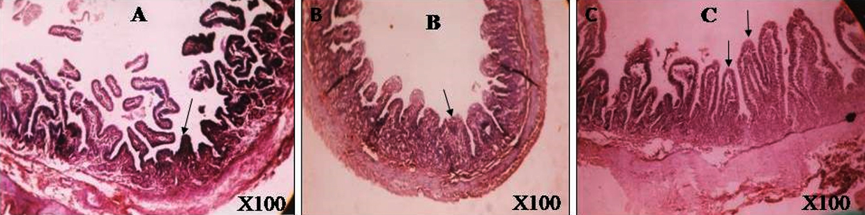
Histological examination: Ileal segments exposed to toxin showed inflammation in mucosa, sub-mucosa and sometimes in the muscle layer. It also revealed enterocytic necrosis and shearing off tip of villi and alteration of villi integrity (Grade-4 inflammation) shown in figure 2A. The section exposed to enterotoxic V. cholerae 569B revealed infiltration of polymorphs in mucosa, sub-mucosa, and muscle layer (Grade-3 inflammation) shown in figure 2B. Villi integrity wasmore or less maintained. No change except mild inflammation was observedin TSB treated loops (Figure 2).
Enterotoxigenic effect by RITARD model:Over a 5-day observation, it was recorded that the rabbit produced mucus containing watery stools within first 24 hours which lasted for 3 days. After five days, an autopsy of the small intestine of rabbits appeared normal.PBS injected rabbits, served as negative controls and did not produce diarrhoeal signs and symptoms.
Neurotoxic effect in mice: LD50 dose of Stx2d was found to be 1 µg toxin(s). The sign of toxic effect appeared within 36 hours which included ruffled fur, huddling and disinclination to move, when effective dose was given. Hind limb paralysis and rapid breathing were observed as signs of severe illness and all mice died within 48 hours. No signs of toxic effect were observed in TSB injected mice.
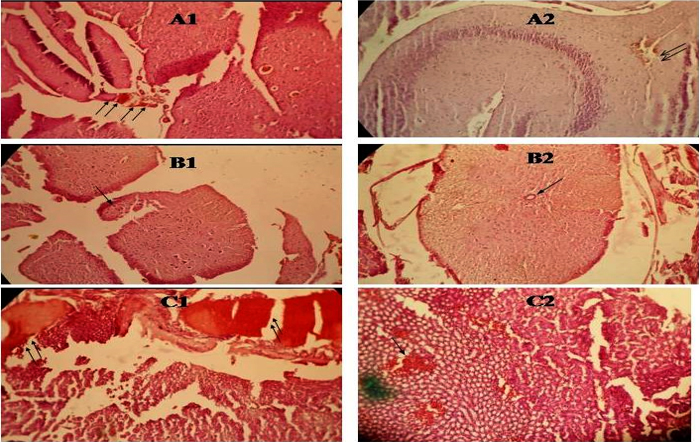
Histological examination: Histological examination of the brain tissue revealed moderate number of congested blood vessels and larger foci of microhaemorrhage (figure 3A1). Spinal cord tissue exhibited moderate number of congested blood vessels, foci of microhaemorrhage and separated cord tissue fragments (figure 3B1). Kidney tissue showed moderate number of congested blood vessels, foci of microhaemorrhage and degenerative to necrotic change in the tubular epithelial cells (figure 3C1). However,TSB treated brain, spinal cord and kidneyshowed unremarkable changes (Figure-3 ).
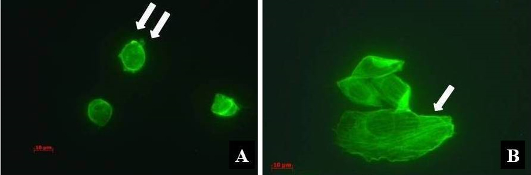
Cytotoxic effectin HeLa cells:Toxin (s) showed cytotoxic potential by studying the morphological changes underepifluorescence microscope,which after 24 hourswas round due to destructionof actin filament as shown in figure 4A.The untreated cells showed normal morphology with actin filament (Figure-4).
Discussion
Patients infected by STEC initially suffer from watery diarrhoea, but in some cases progresses to bloody diarrhoea and haemorrhagic colitis.26-28 The roles of particular genotypic variants in human pathogenesis are not clear. However, Stx2d producing strain has been shown to be the strongest among all variants.4In this study, STEC PT187positive for stx2dstrain was identified from diarrheal patientsand its toxic activities were investigated. Earlier it was reported in 1995 that, STEC infections among the patients with diarrhoea was not identified in Bangladesh.29 Studying the pathogenicity of this strain showed that the toxin(s) has ability to induce fluid accumulation in rabbit ileal loop (figure 1) and cause inflammation and alteration of villi integrity(figure 2). Further evidence showed that this toxin produced mucus-producing diarrhoeain the RITARD model test. STEC strains are a diverse group in their capacity to cause serious diseases in humans, and their key determinants of virulence are their ability to adhere to intestinal epithelial cells and colonize the human gut. Previous studies have shown that shiga toxin causes damage to villus cells by destroying absorptive cells and causing net fluid secretion.30
Previously, it has been reported that, neurological disorders are the most frequent extra-intestinal manifestations of shigellosis occurring in children as well as in adults.31 Convulsions were observed as the most significant neurological symptom of STEC infection.32,33 The findings of Richardson et al., displayed disruption of the nerve supply to the limbs causing paralysis.34 This coincides withresults from this studywhich showed that the toxin(s) caused hind limb paralysis and damage to nervous tissues within mice model (figure3). Obata and his colleagues also reported that Stx2 causes altered neuronal function leading to paralysis.35
Kidney tissue treated with this toxin(s) in mice caused degenerative to necrotic change in the tubular epithelial cells (figure 3). During histopathological observation, damage to the glomerular endothelial cell is referred to as a hallmark of HUS.36 STEC infection progressing to HUS, is characterized by a triad of acute renal failure, microangiopathic haemolyticanaemia, and thrombocytopenia.37 Some individuals with HUS experienced neurological symptoms including lethargy, severe headache, convulsions, and encephalopathy.38 Additionally, the toxin(s) hascytotoxic activity by destroying actin filament in HeLa cell (figure 4). Shiga toxin induced cell death is an important process in the pathophysiological response of humans.
Conclusion
The present study represents functional studies from stx2d gene positive strain, STEC (PT187). The Stx2d produced by STEC showed both enterotoxic and neurotoxic activities. The toxin(s) also exhibited strong cytotoxic potential in animal models. Results from the animal model and human cell studies were found to be consistent with clinical abnormalities shown in human STEC infections.
Acknowledgement
This research study was funded by icddr,b and its donors, which provided unrestricted support to icddr,b for its operations and research. Current donors providing unrestricted support include: Australian Agency for International Development (AusAID), Government of the People’s Republic of Bangladesh; Canadian International Development Agency (CIDA), Swedish International Development Cooperation Agency (Sida), and the Department for International Development, UK (DFID). Authors gratefully acknowledge these donors for their support and their commitment to icddr,b's research-based efforts. The funders had no role in study design, data collection and analysis, decision to publish, or preparation of the manuscript.
References
- Paton AW, Paton JC. Detection and characterization of Shiga toxigenic Escherichia coli by using multiplex PCR assays for stx1, stx2, eaeA, enterohemorrhagic E. coli hlyA, rfbO111, and rfbO157. J. Clin. Microbiol.1998;36:598-602.
Doi: - Islam MA, Heuvelink AE, de Boer E, Sturm PD, Beumer RR, et al. Shiga toxin-producing Escherichia coli isolated from patients with diarrhoea in Bangladesh. J. Med. Microbiol.2007;56:380-385.
Doi: - Tarr PI, Gordon CA, Chandler WL. Shiga toxin-producing Escherichia coli and haemolyticuraemic syndrome. Lancet. 2005;365:1073-1086.
Doi: - Melton-Celsa AR, Darnell SC, O’Brien AD. Activation of Shiga-like toxins by mouse and human intestinal mucus correlates with virulence of enterohemorrhagicEscherichia coli O91:H21 isolates in orally infected, streptomycin-treated mice. Infect. Immun.1996;64:1569-1576.
Doi: - Pierard D, Muyldermans G, Moriau L, et al. Identification of new verocytotoxin type 2 variant B-subunit genes in human and animal Escherichia coli isolates. J. Clin. Microbiol.1998;36:3317-3322.
Doi: - Schmidt H, Scheef J, Morabito S,Stevens D, Lauwers S, et al. A new Shiga toxin variant (Stx2f) from Escherichia coli isolated from pigeons. Appl. Environ. Microbiol.2000;66:1205-1208.
Doi: - Kuczius T, Bielaszewska M, Friedrich AW,Zhang W. A rapid method for the discrimination of genes encoding classical Shiga toxin (Stx) 1 and its variants, Stx1c and Stx1d, in Escherichia coli. Mol. Nutr. Food Res.2004; 48:515-520.
Doi: - Kokai-Kun JF, Melton-Celsa AR,O’Brien AD. Elastase in intestinal mucus enhances the cytotoxicity of Shiga Toxin type 2d. J. Biol. Chem. 2000;275:3713-3721.
Doi: - Tasara T, Bielaszewska M, Nitzsche S,Karch H, Zweifel C, et al. Activatable Shiga toxin 2d (Stx2d) in STEC strains isolated from cattle and sheep at slaughter. Vet. Microbiol. 2008;131:199-204.
Doi: - Wiley RG, Donohue-Rolfe A,Keusch GT. Axonally transported shigellacytotoxin is neuronotoxic. J. Neuropathol. Exp. Neurol. 1985;44:496-506.
Doi: - World Health Organization. Program for control of diarrheal diseases. In Manual for Laboratory investigation of Acute Enteric Infections, CDD/93.3, rev.1. World Health Organization, Geneva, Switzerland. 1987; P.9-20.
Doi: - Schmidt H, Geitz, Torr PI, Frosch M, Karch H, et al. Non-O157:H7 pathogenic shiga toxin-producing Escherichia coli: phenotypic and genetic profiling of virulence traits and evidence for clonality. J. Infect. Dis.1999;179:115-123.
Doi: - Bonnet R, Souweine B, Gauthier G, Rich C, Livrelli V, et al. Non-O157:H7 Stx2-producing Escherichia coli strains associated with sporadic cases of hemolytic-uremic syndrome in adults. J. Clin. Microbiol. 1998; 36:1777-1780.
Doi: - Karch HH, Huppertz J, Bockemithl H, Schmidt A, Schwarzkopf, et al. Shiga toxin-producing Escherichia coli infections in Germany. J. Food Prot. 1997; 60:1454-1457.
Doi: - Franke S, Gunzer F, Wieler LH, Baljer G, Karch H. Construction of recombinant Shiga-like toxin-IIv(SLT-IIv) and its use in monitoring the SLT-IIv antibody status in pigs. Vet. Microbiol. 1995; 43:41-52.
Doi: - Friedrch A, BielaszewskaM, Zhang WL, Pulz M, Kuczius T, et al. Escherichia coli harbouring Shiga toxin 2 gene variants: frequency anf association with clinical symtoms. J. Infect. Dis.2002;185:74-84.
Doi: - Talukder KA, Azmi IJ, Ahmed KA, Hossain MS, Kabir Y, Cravioto A, Sack DA, Nur-E-Kamal A. Activation of p53/ATM-dependent DNA damage signaling pathway by shiga toxin in mammalian cells.Microb. Pathog. 2012;52(6):311-317.
Doi: - Sanyal, SC, Saraswathi B, Sharma P.Enteropathogenicity of Plesiomonasshigelloides.J. Med. Microbiol.1980;13:401-409.
Doi: - Nur-E-Kamal A, Li TK, Zhang A, Qi H, Hars ES, Liu LF. Single-stranded DNA induces ataxia telangiectasia mutant (ATM)/p53-dependent DNA damage and apoptotic signals. J Biol Chem. 2003; 278:12475-12481.
Doi: - Singh SJ, Sanyal SC. Enterotoxicity of the so-called NAG vibrio. Ann. Soc. Belg. Med. Top. 1978; 58:133-140.
Doi: - Fernandez A, Sninsky CA, O’Brien AD, et al. Purified Shigella enterotoxin does not alter intestinal motility. Infect. Immun. 1984; 43:477-481.
Doi: - Spira MW, Bradley sack R, Froehlich JL, Clench MH, Mathias JR. Simple adult rabbit model for Vibrio cholerae and enterotoxigenicEcherichia coli diarrhea. Infect. Immun. 1981; 32:739-747.
Doi: - MacLeod DL, Gyles CL. Purification and characterization of an Escherichia coli. Infection and Immunity. 1990; 58:1232-1239.
Doi: - Elwell C, Chao K, Patel K, Dreyfus L. Escherichia coliCdtB mediates cytolethal distending toxin cell cycle arrest. Infect Immun. 2001; 69: 3418-3422.
Doi: - Aragon V, Chao K, Dreyfus LA. Effect of cytolethal distending toxin on F-actin assembly and cell division in Chinese hamster ovary cells. Infect. Immun. 1997; 65:3774-3780.
Doi: - O'Brien AD, Lively TA, Chen M, Rothman SW,Formal SB. Escherichia coli O157:H7 strains associated with haemorrhagic colitis in the United States produce aShigelladysenteriae1 (Shiga) like cytotoxin. Lancet.1983;1:702.
Doi: - Riley LW. The epidemiologic, clinical and microbiological features of hemorrhagic colitis. Annu. Rev. Microbiol.1987;41:383-407.
Doi: - Henry FJ, Udoy AS, Wanke CA,Aziz KM. Epidemiology of persistent diarrhea and etiologic agents in Mirzapur, Bangladesh. Acta Paediatr.1996;381:27-31.
Doi: - Albert MJ, Faruque SM, Faruque AS, Neogi PK, Ansaruzzaman M, et al. Controlled study of Escherichia coli diarrheal infections in Bangladesh children. J. Clin. Microbiol.1995;33:973-977.
Doi: - Keenan KP, Sharpnack DD, Collins H, Formal SB, O'Brien AD.Morphologic evaluation of the effects of Shiga toxin and E. coli Shiga-like toxin on the rabbit intestine. Am. J. Pathol. 1986;125:69.
Doi: - Zajdowicz T. Epidemiologic and clinical aspects of shigellosis in American forces deployed to Saudi Arabia. South Med. J.1993;86:647-650.
Doi: - Kowlessar M, Forbes GB. The febrile convulsion in shigellosis. N. Eng. J. Med.1958;1258:520-526.
Doi: - Avital A, Maayan C, Goitein KL. Incidence of convulsions and encephalopathy in childhood Shigella infections. Clin. Pediatr. (Phila). 1982;21:645-648.
Doi: - Richardson SE, Rotman TA, Jay V, Smith CR, Becker LE, et al. Experimental verocytotoxemia in rabbits. Infect. Immun.1992;60:4154-4167.
Doi: - Obata F, TohyamaK, Boney AD, Kolling GL, Keepers TR, et al. Shiga toxin 2 affects the central nervous system through receptor globotriaosyceramide localized to neurons. J. infect. Dis.2008;198:1398-1406.
Doi: - Fontaine A, Arondel J, Sansonetti PJ. Role of Shiga toxin in the pathogenesis of bacillary dysentery, studied by using a Tox-mutant of Shigelladysenteriae 1. Infect. Immun.1988;56:3099-3109.
Doi: - Karmali MA, Petric M, Lim C, Fleming PC, Arbus GS, et al. The association between hemolytic uremic syndrome and infection by Verotoxin-producing Escherichia coli.J. Infect. Dis. 1985;151:775-782.
Doi: - TeshVL, O’Brien AD. The pathogenic mechanisms of Shiga toxin and the Shiga-like toxins. Mol. Microbiol. 1991;5:1817-1822.
Doi:
Departemnt of Microbiology, University of Dhaka, Bangaldesh.
crahsan@du.ac.bd
 0000-0001-5024-6293
Submission
2019-10-29
Accepted
2020-03-01
Published
2020-04-01
Apply citation style format of Bangladesh Medical Research Council
Issue
Vol 46 No 1 (2020)
Section
Research Articles
Financial Support
None
Conflict of Interest
The authors declare no conflict of interest.


